An ultrasound is an evaluation tool commonly used by doctors during pregnancy.
It is a safe and useful way to obtain images of the baby and its environment to provide useful information about your health.
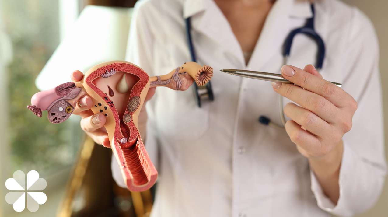
Ultrasounds in pregnancy during 6 and 12 weeks
The first ultrasound is recommended in the first trimester of pregnancy. In a typical situation, the scan will show the early pregnancy sac in the uterus with the fetus inside the sac. This can be seen from 5 to 6 weeks of pregnancy. Visualizing the pregnancy in the womb is reassuring, because it confirms that it is in the right place.
Although very rare, the pregnancy can sometimes implant in the wrong place, such as in the fallopian tubes (known as an ectopic pregnancy). This happens to 1-2% of all pregnancies.
The baby's heartbeat is normally visible at 6 weeks of pregnancy. Sometimes the doctor may use a vaginal probe for the examination when the pregnancy is between 5 and 6 weeks. The vaginal scan is safe and does not harm the pregnancy in any way.
The current gestation (age) of the pregnancy can be estimated by taking measurements of the baby during the scan. The due date can be estimated accurately. This is especially important for the doctor to monitor the pregnancy well.
It also helps with planning ahead, such as estimating time off work and making all necessary arrangements for baby care. In fact, the accuracy of first trimester ultrasound is within a week. The ultrasound of the first trimester also allows to know if the quantity of implanted embryos.
Nuchal translucency scan: weeks 11 to 14
In recent years, first-trimester ultrasounds can be used to assess your baby's risk of being affected by Down syndrome.
This is done by measuring the thickness of the skin fold (nuchal translucency) at the back of the baby's neck. If the neck crease is unusually thick, it may indicate that the baby may have Down syndrome.
Other causes may include heart abnormalities or even rarer genetic syndromes. This test is quite accurate in detecting Down Syndrome as the detection rate is 80% in the hands of experienced doctors.
If your baby is in an optimal position, the ultrasound exam after 11 weeks can visualize the bone of the baby's nose (known as the nasal bone). The absence of the nose bone is a worrying sign, which increases the risk of Down syndrome.
Screening scan: week 18 to 22
An ultrasound can be used to detect abnormalities in the physical structure of the baby, for example, in the heart, lungs, spine, brain, long bones of the legs or arms, organs in the abdomen such as the liver, the stomach, intestines, kidneys, bladder and even check for cleft lip and palate.
The finding of certain abnormalities during an ultrasound can also alert doctors to the possibility of Down syndrome or other genetic abnormalities in the baby. Further tests can then be done to exclude them.
Ultrasound screening for physical abnormalities is usually done around the fifth month or 20th week of pregnancy. The accuracy of the scans is around 70% in detecting all anomalies.
Growth scan: 32 to 36 weeks
Later in the pregnancy, it is important to monitor the growth of the baby. The doctor will monitor this, most of the time by examining and measuring the height of the belly during visits.
Ultrasound in the late second trimester can help confirm that the baby is growing well. You can also tell if the amniotic fluid in the "bag of waters" is enough for the baby. This tells us about the baby's health status.
doppler scan
Sometimes a more specialized mode of ultrasound called Doppler ultrasound may be used for more specific conditions. For example, when the growth of the baby is reduced or anemia (low hemoglobin) is suspected in the baby. Doppler scanning can help provide additional information about the status of placental blood flow and the health status of the baby.
El Dr. Daniel García, gynecologist and obstetrician at Ginefem, explains the types of existing ultrasounds for pregnant women.